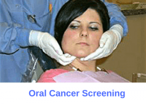
Oral cancer accounts for 1% of all cancers worldwide. While more prevalent in smokers, oral cancer can certainly develop in non-smokers as well, regardless of your age. Like most other cancers, the key to successful treatment is early diagnosis. Dentists spend a lot of time looking in your mouth. This provides them an excellent opportunity to screen for oral cancer lesions. A comprehensive oral cancer screening can detect early symptoms of cancer while still in a treatable state.

Oral cancer screening requires visual and manual examination
Oral Cancer Screening
Oral cancer screening should be a part of every new dental patient examination. A comprehensive oral cancer screening consists of both a visual and a manual exam.
Visual Screening
Your dentist will look for any suspicious-looking lesions. First, they examine your head and neck region. Next, they evaluate the inside of your mouth which includes your cheeks, tongue, palate, and gum tissue. Cancerous and pre-cancerous lesions typically appear as red or white spots inside the mouth. These lesions typically have undefined, raised borders, and occasionally demonstrate ulceration. If your dentist identifies a suspicious lesion they will take further action.
Manual Exam
Manual oral cancer screening involves palpation of your head and neck. Oral cancer travels through the head and neck lymph nodes, therefore it's important to examine this area closely. Suspicious nodes typically manifest themselves as firm and non-tender nodules. Your dentist will palpate your occipital, postauricular, preauricular, anterior cervical, posterior cervical, supraclavicular, submandibular, and submental lymph nodes, basically your entire head and neck region, to look for suspicious lymph nodes.
What happens if my dentist detects a suspicious lesion?
Should your dentist finds a suspicious-looking lesion or node, they will make a note of it and inform you of their findings. Based on the appearance of the lesion, its location, and your risk levels, your dentist may recommend one of the following:
Follow-up
A follow-up is recommended if your lesion doesn't appear too suspicious and you're a low-risk patient. You will be scheduled for a follow-up typically in two to three weeks. Should the same lesion or nodule still persist at this time, then further action is warranted.
Biopsy
There are a few different ways to perform a biopsy of an oral lesion. One option is to perform a brush biopsy. A brush biopsy is a painless and simple procedure that requires no anesthesia, cutting, or drilling of the suspicious lesion. The other option is an excisional biopsy. This is where your dentist or oral surgeon cuts the lesion and sends it to the pathologist for a biopsy.
Referral
Sometimes your dentist may refer you to an appropriate specialist for further evaluation. This may include an oral surgeon, dermatologist, or other physicians. Be sure to schedule your appointment and inform your dentist of their findings.