Why are X-rays important in dentistry?
Dentists rely heavily on X-rays to diagnose hidden dental problems. Dental cavities, gum disease, infections, and many other oral lesions can only be detected in their early stages by taking proper radiographs. Without X-rays, your dentist won't be able to diagnose cavities until they cause pain. Early-stage gum disease can be detected by looking at X-rays of your teeth. Some oral infections don't manifest any symptoms and can only be diagnosed through X-rays. Dentists also use X-rays to communicate with specialists, insurance companies, and other dental providers. Taking X-rays is an essential part of dentistry, which is why most dental visits start off by taking X-rays of your teeth.

X-rays are important to diagnosing most oral conditions
What are some common X-rays used in dentistry?
There are about three to four common types of X-rays used in dentistry. Dentists use different X-rays for different reasons. Some X-rays are used to diagnose dental cavities and gum disease. Some X-rays are used to visualize nerve canals and sinus membranes. Others X-rays are used for orthodontic diagnosis. Here are the most common types of X-rays used to diagnose various oral conditions:
- Periapical X-rays & bitewing X-rays
- Panoramic radiograph
- Lateral cephalometric
- CT/CBCT scan
Pericapical & Bitewing X-rays
Periapical and bitewing X-rays are those little X-rays that the dental assistant takes while you sit in the dental chair. Open, close, bzzzz! Periapical and bitewing X-rays capture lots of fine details on your teeth. Therefore, they are ideal X-rays for diagnosing dental cavities, tooth infections, and even gum disease. Most periapical and bitewing X-rays cover three, four, or maybe five teeth. You need about a dozen or so X-rays to capture the whole mouth. Most dentists typically take anywhere from 12 to 14 periapical X-rays as well as 4 bitewing X-rays on your initial visit. This is referred to as full mouth X-ray series. These X-rays provide your dentist with a full view of what your teeth and jawbone look like. These X-rays are followed by an additional 6 to 8 radiographs on future recall appointments. Recall X-rays are used to diagnose new issues that your teeth may be facing.
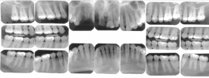
Periapical and bitewing X-rays show plenty of details, but each X-ray only captures a small region of the mouth
Panoramic X-ray (Panorex)
Panoramic radiographs are used to visualize wisdom teeth, impacted teeth, sinuses, as well as major nerves and arteries. They also assist with diagnosing Temporomandibular Joint (TM) problems. These X-rays capture a large segment of your skull. They provide your dentist with a broad overview of what your teeth, jaws, and facial bones look like. Panoramic X-rays are useful for oral surgery procedures like tooth extraction and dental implant placement. However, they are not useful for diagnosing dental cavities. While very useful for many procedures, Panoramic X-rays are not a substitute to traditional radiographs for diagnosing cavities.
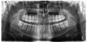
Panoramic X-rays capture the wisdom teeth, Inferior Alveolar nerve & sinus membranes
Lateral Cephalometric
Lateral cephalometric X-rays are primarily used for orthodontic diagnosis. These X-rays show the relationship between your upper and lower teeth and jawbone from a lateral perspective. Orthodontists use lateral cephalometric X-rays to identify your jaw relationship and monitor your braces treatment. Lateral Cephalometric X-rays can also be used for the diagnosing obstructive sleep apnea (OSA) and certain ENT conditions.
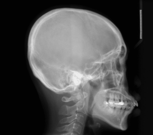
Lateral cephalometric X-ray shows the relationship between the upper and lower teeth and jaws
Computed Tomography (CT Scan)/ Cone Beam Computed Tomography (CBCT Scan)
CT scans and CBCT scans capture 3-D images of your facial structures. CT scans are used to capture a 3-D image of the whole jaw. CBCT scans can only capture a small region and not the entire skull. Because CT scans and CBCT scans are 3-D images, they capture details that go undiagnosed on regular X-rays. The main use of CT and CBCT images is to identify important landmarks during dental implant surgery. These X-rays are used to measure bone width, identify nerve location, and measure the jawbone under your sinus membranes. In fact, a CT or CBCT scan is the only acceptable diagnostic radiograph for treatment planning implant dentistry.
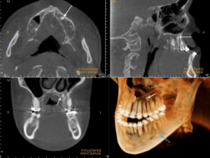
CT scans and CBCT scans capture 3-D views of your skulls
Which X-rays do I need?
It all depends on which dental treatment you need. Periapical X-rays and bitewing X-rays are the most commonly used types of dental X-rays. If you need a couple of cavities fixed or dental cleaning, then you probably need to take a few bitewing and periapical X-rays. If you need your wisdom tooth removed then you need a panoramic X-ray, possibly a CT scan. Braces treatment for overbites and underbites may require a lateral cephalometric X-ray. Dental implants usually require a CT or CBCT scan and other necessary radiographs. Discuss your issues with your dentist to determine which X-rays are the most appropriate for your diagnosis.